Neuroscan Curry Multi-Modal Neuroimaging
Multi-Modal Neuroimaging
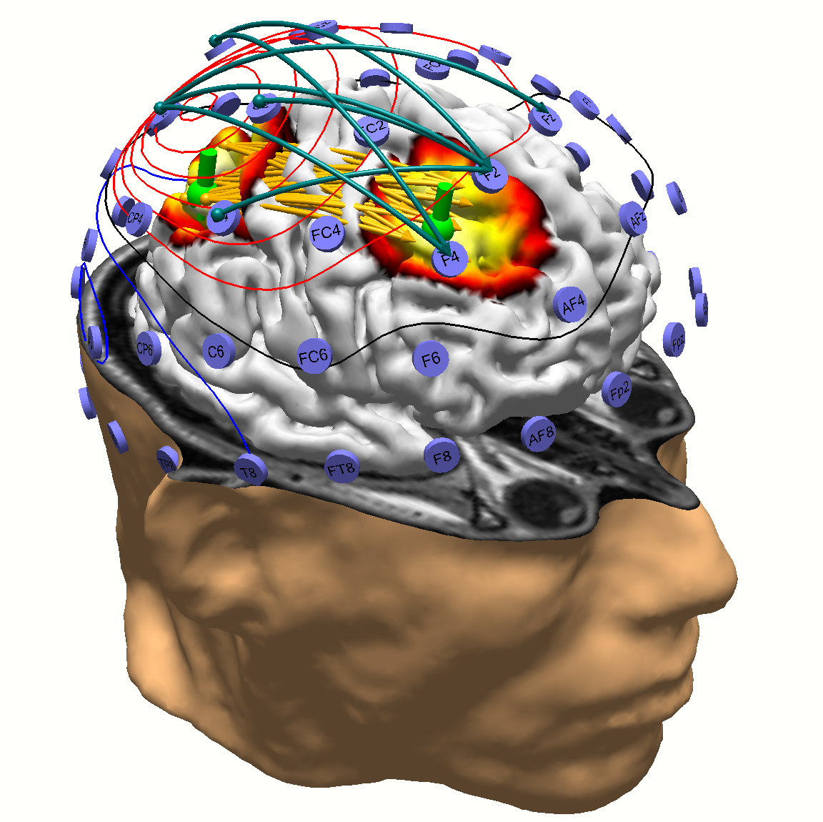
CURRY integrates multiple complementary functional and imagemodalities (EEG, ECoG, MEG; MRI, fMRI, PET, SPECT and CT) in a single software package for the purpose of obtaining the maximum accuracy of electrical source analysis. CURRY uses the full physical anatomy from MR and CT to provide three-dimensional models of the skull and brain allowing the neural generators of the topographic EEG to be computed.
CURRY was developed for mapping and identification of the neural generators of EEG and MEG recordings. However, the functionality offered and the revolutionary changes in this latest version CURRY also make it suitable for wider application, including clinical applications in neurology, epilepsy and radiology.
• Integration of EEG, MEG, ECoG, ECG, MCG, with MRI, fMRI, CT, PET, SPECT.
• Complete data processing from filtering to source analysis.
• Event support, template-based event detection.
• Principal and Independent Component Analysis (PCA, ICA) and filtering.
• Individual realistic head models using the Boundary Element Method (BEM).
• Pre-computed BEM and Finite Element Method (FEM) head models.
• Dipole fits. Dipole confidence ellipsoids are computed.
• Dipole scans, extended source (patch) scans, and MUSIC scans.
• Beamforming based on dipolar or extended sources.
• Current density analysis, extended sources, Lp norms, sLORETA, SWARM.
• Export of results in Excel, MATLAB, and SPM formats.
• Report generator.
• Windows multi-document user interface.
• Data import wizards for functional and image data.
• Multi-core support using thread-based multitasking and parallelization.
• Hardware accelerated real-time rendering of 3D scenarios.
• Context-sensitive HTML help system.
• FDA Market Clearance for intended clinical applications.
• Online updates
Curry 8 impovements
FOR ALL USERS
- New, easy-to-use and customizable User Interface
- Customizable function keys (F-keys) for quick selection of montages or other actions
- No memory limits (native 64-bit application)
- Interpolate any surface EEG into 10-20 system(or other) with the click of one button
- Automatic Hi gh-Tesla MRI histogram correction
- Exported data files contain a file history, documenting previous filenames and processing steps
- Automatic creation of individual Finite Element Models (FEM)
FOR CLINICIANS
- Easy dipole clustering
- Simultaneous display of five image data sets (full support for up to ten image data sets)
- Surgical navigation systems: RGB DICOM
- Source reconstruction on depth electrodes (stereo-EEG) is now possible
- Workflows with Scope(*) support (*) Scopes are sets of factory defaults and macros for certain application areas. Epilepsy, ERP, and CURRY 7 scopes are provided; custom scopes can be created
FOR RESEARCHERS
- Faster digitization of electrode positions
- Flexible signal processing sequences
- New statistics on source reconstruction results and channels
- New wavelet and Current Density Reconstruction (CDR) options
- Improved automation: macros with jumps, loops, branches, subroutines
- Stream live data to MATLAB or via TCP/IP (NetStreaming), dedicated to Brain-Computer-Interface (BCI) applications
- Data files compatible with MATLAB and EEGlab


